Inside the Ko Lab
Tracking Living Tissues
The Challenge
When scientists create methods to detect disease biomarkers, they give healthcare providers better tools to properly diagnose and treat patients. However, limitations to obtaining this information, especially when using living cells and tissues from patients, prevents a complete picture of what is unique about a case and decreases the chance that the best course of treatment can be identified.
The Status Quo
Several techniques exist for identifying multiple biomarkers in cells, but they are usually not compatible with observing changes over time in living cells or are limited by a set number of biomarkers that can be profiled. The chemicals used to profile multiple (>5) biomarkers are toxic to the cells, preventing live cell monitoring. Due to this limitation, a full understanding of the protein expressions of the living cells could not be obtained and a clear picture of what is actually occurring during the course of cellular changes was out of reach.

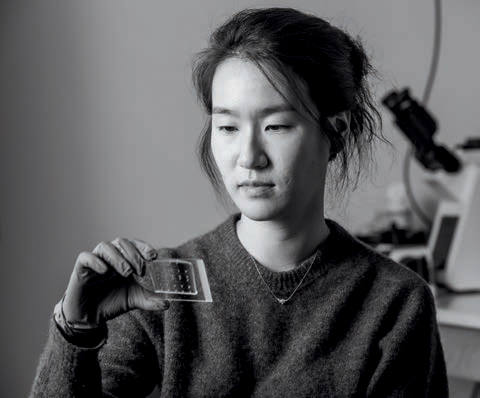
Jina Ko
Assistant Professor in Bioengineering, Penn Engineering;
Pathology and Laboratory Medicine, Perelman School of Medicine
The Ko Lab’s Fix
Jina Ko, Assistant Professor in Bioengineering, is working to overcome this limitation with a method known as “scission-accelerated fluorophore exchange” (or SAFE), a new way to detect biomarkers in cells that is highly gentle and allows for high multiplexing via cyclic imaging so that more biomarkers can be identified in a single sample and changes in living cells and tissues can be tracked over time. She first developed this method during her postdoctoral training at Massachusetts General Hospital under the supervision of Jonathan Carlson and Ralph Weissleder.
The method uses “click” chemistry, which is a bioorthogonal, non-toxic and rapid reaction that allows the team to highlight the desired biomarkers in the samples without destroying them each time a microscopy cycle is run.
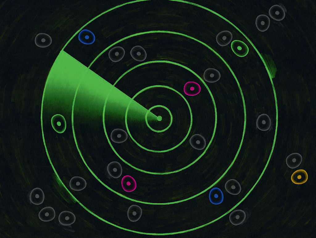
SAFE can track nearly as many markers as necessary with ultra-fast kinetics because researchers can repeatedly stain cells in a living sample using the developed probes with fluorophores and then release those fluorophores following the imaging, repeating this cycle multiple times to achieve multiplexing. A cycle can profile 3 biomarkers, so if a researcher needs information on 15 markers, they would run the cycle 5 times to create the cell profile that they need within an hour.
Understanding activity in an individual’s living samples over time addresses the issue of subtlety and heterogeneity when identifying a patient’s unique disease response and allows for more tailored treatment approaches.
“You can’t identify a treatment that works for the average person, apply that treatment to everyone and expect the best outcomes,” says Ko. “Using this method, if we want to administer a therapy to a patient, we could remove a sample of their cells and use that sample to try different therapeutic options. After tracking the sample, we could predict if the patient would respond well to therapy A, but not therapy B. Our goal is that this technology will be applied in the clinic to help patients.”
Text by Olivia McMahon / Photo by Kevin Monko/ Illustration by Pete Ryan
